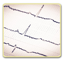
Last week’s Health Tip discussed the usefulness of the EKG, a highly important test used in the diagnosis and management of heart disease. Like most clinical tests, however, the EKG is not perfect. Some of the ways in which the EKG falls short in evaluating heart conditions include:
- Evaluating intermittent problems. Most of us know how difficult it can be to have a problem that comes and goes affecting an automobile “diagnosed” by a mechanic. The same can be true with an intermittent problem affecting the heart, such as palpitations. Unless the person with the irregular heart beat is experiencing symptoms or signs of the condition at the time that the EKG is being performed, it will most likely appear normal.
- Evaluating heart problems that occur only with activity. The EKG looks at the electrical activity of the heart only in a resting or “static” state. Underlying heart problems may not be reflected unless the heart is beating rapidly or is under stress as when exercising. An example of this is someone with constriction of the arteries that supply oxygen to the heart (coronary artery disease) who experiences chest pain (angina) with activity. Often, when this person is at rest, his or her EKG will not reflect the underlying heart condition.
- Producing false-positive and false-negative findings. Sometimes the EKG can be overly sensitive and point toward a heart problem that is not really present. This is called a “false-positive” finding. Likewise, an EKG may be entirely normal despite the presence of a serious underlying heart condition. This is called a false-negative finding and is a likely explanation for the person who has a normal EKG during a routine physical exam and shortly afterwards experiences a serious heart attack.
- Causing non-specific changes on EKG. Sometimes the lines and waves seen on an EKG are “non-specific”, meaning that they may or may not be abnormal. Some of these findings are known as “normal variants”, appearing abnormal but occurring without the presence of heart disease. Other non-specific changes could be due to a number of conditions without pointing to a specific cause.
- Erroneous diagnosis of heart disease in athletes. As a result of superb conditioning and physiologic adaptation to exercise, athletes may have EKG “abnormalities” that pose no health risk. A slow heart rate (sometimes as low as 40 beats/minute), enlargement of the heart similar to that seen in long-standing hypertension, and changes in electrical activity (ST segment elevation, inverted T-waves, etc.) suggesting heart stress are some of the most common of these findings.

How can these limitations of the EKG be overcome? When the EKG findings and the patient’s physical condition are at odds, a more in-depth examination, and often other heart tests, may be required to sort things out. The following are some of the most common of these tests:
- Ambulatory electrocardiography. In this test, also known as a 24-hr EKG or Holter monitor, a 24 hour recording of the EKG is made while you are going about your normal activities. It is particularly valuable in someone who has an intermittent problem, such as palpitations. Also, some heart problems are only present during activity which an ambulatory electrocardiogram will help document.
- Echocardiography. The echocardiogram uses sound waves to scan the heart’s muscle and valves that control the flow of blood through and out of the heart. It even allows doctors to see the heart while it is beating. The echocardiogram is especially valuable in and in assessing abnormal heart sounds heard during examination, evaluating an enlarged heart, and in helping clear athletes with questionable EKG changes.
- Exercise electrocardiography. This test is also known as a “treadmill EKG” or “stress test”. During this test the EKG is monitored while the person is walking on a motor-powered treadmill or pedaling a stationary bicycle. Exercise often provides the additional stress on the heart required to bring out EKG evidence of coronary artery disease.
On occasion even more invasive testing such as a coronary artery catheterization may be required to confirm or rule out a problem noted on EKG. This is part of the reason that the US Preventive Services Task Force has advised against doctors performing “screening” EKGs on people at low risk of having heart disease. In “normal” individuals, the EKG is unlikely to predict a heart attack or uncover an undiagnosed heart problem, and because of false-positives and non-specific findings may lead to unnecessary testing.
